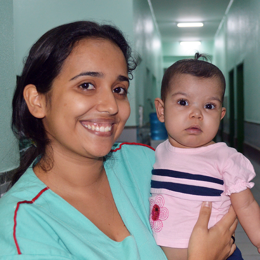
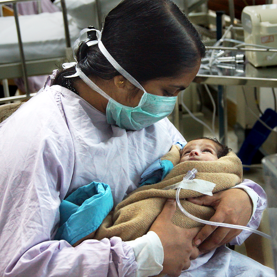
Congenital Heart Disease Around the World
One out of 100 babies is born with congenital heart disease (CHD). This is heart disease due to an abnormality in the structure of the heart. A child born in an area with access to pediatric heart care and surgery will likely survive and go on to live a healthy and active life. Sadly, for 9 in 10 of these children, treatment is not available or too expensive. Children’s HeartLink is working to change that by training in-country pediatric heart teams to treat children in Bangladesh, Brazil, China, India, Malaysia and Vietnam.
Congenital Heart Disease Facts
- CHDs are the leading cause of infant illness and death from birth defects. Read our findings in the first Global Burden of Congenital Heart Disease Study in The Lancet Child & Adolescent Health.
- The survival of infants with CHD depends on how severe the defect is, when it is diagnosed and how it is treated.
- About 1 in 4 of babies with CHDs have a critical CHD. These infants generally need surgery or other procedures in their first year of life. In general, survival and medical care for babies with critical CHD are improving.
- At least 15% of CHDs are associated with genetic conditions.

Access to Care in Countries Where We Work
- In India, only one in 10 children with congenital heart disease receives optimal care. The limited number of pediatric heart care programs and uneven geographic access to these services are among the reasons. Read about our work in India
- Access to care for a child with heart disease born in rural China is almost nonexistent. Read about our work in China
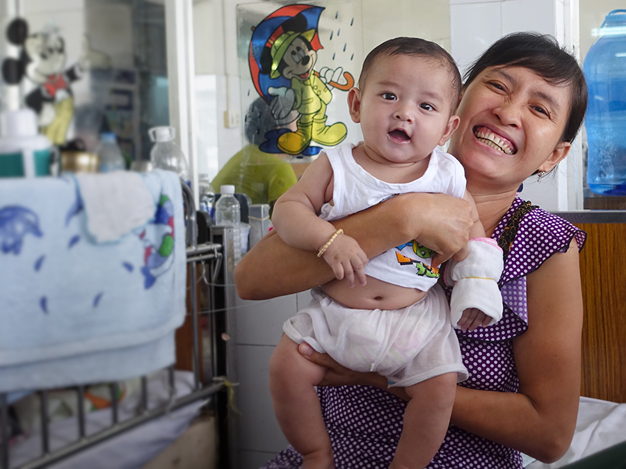
- In Vietnam, there are only 20 pediatric cardiologists to serve the 50,000 children in need. Read about our work in Vietnam
- Only one-third of children born with heart disease in Brazil have access to treatment. Read about our work in Brazil
- An estimated 3,000 children are born with CHD every year in Malaysia, but not all children receive the timely care they need. Read about our work in Malaysia
Common Types of Congenital Heart Defects
Atrial Septal Defect (ASD)
A hole in the wall (septum) between the two upper chambers of the heart (atria). The hole increases the amount of blood that flows through the lungs. Over time, this may cause problems, such as high blood pressure and even heart failure. A hole can vary in size and may close on its own during infancy or early childhood, or the child may need surgery.
Atrioventricular Canal Defect
A large hole in the center of the heart affecting all four chambers. Valves don’t properly route the blood to each station of circulation. An atrioventricular canal defect allows the oxygen-poor and oxygen-rich blood to mix. This results in extra blood flow to the lungs that forces the heart and lungs to work extra hard. It may lead to heart failure and high blood pressure in the lungs. Surgery is needed to close the hole and reconstruct the valves. This defect is also called an atrioventricular septal defect (AVSD).
Coarctation of the Aorta (CoA)
The major artery that carries blood to the body is narrower than it should be. This narrowing affects oxygen-rich blood flow to the upper and lower parts of the body. If the aorta is not widened, it can cause high blood pressure or heart damage.
Ebstein Anomaly
A tricuspid valve between the two right chambers sits lower than normal and is malformed. Blood may leak backward into the right atrium, making the heart work less efficiently. Ebstein Anomaly may lead to enlargement of the heart or heart failure. Treatment options include medications and surgery.
Hypoplastic Left Heart Syndrome (HLHS)
Babies with HLHS are born with an undeveloped left side of the heart. They have a very small left ventricle, the lower chamber that pumps blood into the aorta. The mitral and aortic valves are very small or completely closed. The mitral valve separates the left ventricle and the left atrium. The aortic valve separates the left ventricle and the aorta. Three surgeries during infancy and early childhood help make the right ventricle the main pumping chamber to the body.
Patent Ductus Arteriosus (PDA)
A hole between the two major blood vessels leading from the heart, the pulmonary artery and the aorta. A small hole often doesn’t cause problems and might never need treatment. A large hole needs to be closed, as it allows poorly oxygenated blood to flow in the wrong direction. Otherwise, it may weaken the heart muscle and cause heart failure.
Pulmonary Artesia
The pulmonary valve is nonexistent. This valve is needed to control blood flow from the lower right chamber (ventricle) to the main pulmonary artery. This artery carries blood from the heart to the lungs. As a result, no blood can flow from the right ventricle to the lungs. In most cases, a baby needs surgery right after birth.
Pulmonary Valve Stenosis
A thickened or fused pulmonary valve that is not fully open. Located between the right heart’s chambers, this valve allows blood to flow into the pulmonary artery and then to the lungs. Pulmonary valve stenosis ranges from mild, without symptoms, to severe cases that require treatment.
Tetralogy of Fallot
A combination of four heart defects. It includes a ventricular septal defect, a hole between the lower two chambers (ventricles). It also includes pulmonary valve stenosis, a narrowing of the pulmonary valve that reduces blood flow to the lungs. Additionally, the aorta, the main artery leading out to the body, lies over the hole in the ventricles. Finally, the muscular wall of the right ventricle is thicker than normal. All babies who have Tetralogy of Fallot need corrective surgery.
Total Anomalous Pulmonary Venous Return (TAPVR)
The pulmonary veins don’t connect to the left upper chamber (atrium). These veins are needed to bring blood back from the lungs. As a result, the oxygen-rich blood returns to the right side of the heart. There it mixes with oxygen-poor blood. This causes the baby to get less oxygen than needed. Babies with TAPVR need surgery to repair the defect.
Transposition of the Great Arteries (TGA)
Two main arteries – the main pulmonary artery and the aorta – connect to the wrong chambers. In a healthy heart, the right side pumps oxygen-poor blood to the lungs through the pulmonary artery. In babies with TGA, oxygen-poor blood enters the right side and is pumped back out through the aorta. The left side of the healthy heart pumps oxygen-rich blood to the body through the aorta. In the TGA heart, oxygen-rich blood from the lungs is pumped back to the lungs through the main pulmonary artery. All babies born with TGA need surgery.
Tricuspid Artesia
There is no tricuspid valve, which is needed to control blood flow from the right upper chamber (atrium) to the right lower chamber (ventricle). As a result, blood is unable to flow to the right ventricle and out to the lungs. For this reason, the right ventricle can be underdeveloped. Surgical treatment will help restore heart function.
Truncus Arteriosus
There is only one blood vessel coming out of the heart. There should be two: the main pulmonary artery and the aorta. In babies with a truncus arteriosus, oxygen-poor blood and oxygen-rich blood are mixed as blood flows to the lungs and the rest of the body. As a result, too much blood goes to the lungs and the heart works harder to pump blood to the rest of the body. Surgery is usually needed in the first few months of life.
Ventricular Septal Defect (VSD)
A hole in the wall (septum) between the two lower chambers (ventricles). The wall keeps the oxygen-rich blood from mixing with the oxygen-poor blood. Many VSDs are small and close on their own. Babies with a large VSD may need surgery. When the hole does not close, it may increase pressure in the heart, reduced oxygen to the body or even heart failure.
Symptoms of a CHD
According to Mayo Clinic, serious congenital heart defects usually become evident soon after birth or during the first few months of life. Signs and symptoms could include:
- Pale grey or blue skin color (cyanosis)
- Rapid breathing
- Swelling in the legs, abdomen or around the eyes
- Shortness of breath during feedings, leading to poor weight gain
Less serious congenital heart defects may not be diagnosed until later in childhood because a child may not have any noticeable signs of a problem. If signs and symptoms are evident in older children, they may include:
- Easily becoming short of breath during exercise or activity
- Easily tiring during exercise or activity
- Fainting during exercise or activity
- Swelling in the hands, ankles or feet